
For 11 Organo Chlorine pesticides Rs.10,500/-+GST per sample
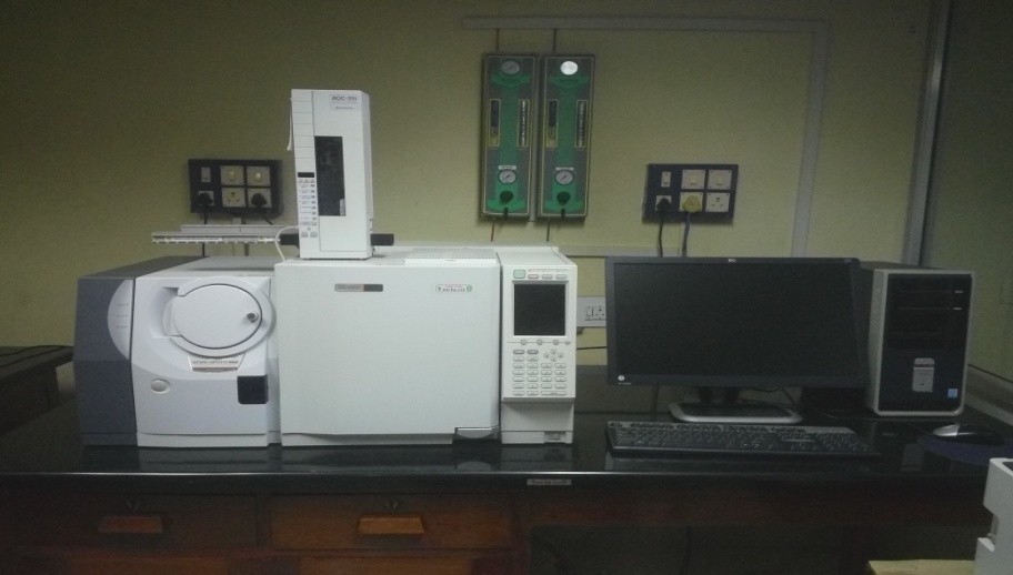
Make: SHIMADZU, JAPANGC-MS QP 2010Plus, 2009
Specification:
Linear temperature elevation rate: 70°C /min up to 200°C
50°C /min up to 300°C
30°C /min up to 400°C
:Injection port temperature: Max 450°C
Temperature: 100-300°C
Filament: Dual filament, Automatic Switching
Electron Voltage Setting range: 10-200 V
Electron current setting range: 5-250µA
Accessories
Description
The Gas Chromatograph–Mass Spectrometer (GC-MS) is composed of two major building blocks: the gas chromatograph and the mass spectrometer. The gas chromatograph utilizes a capillary column which depends on the column's dimensions (length, diameter, film thickness) as well as the phase properties (e.g. 5% phenyl polysiloxane). The difference in the chemical properties between different molecules in a mixture and their relative affinity for the stationary phase of the column will promote separation of the molecules as the sample travels the length of the column. The molecules are retained by the column and then elute (come off) from the column at different times (called the retention time), and this allows the mass spectrometer downstream to capture, ionize, accelerate, deflect, and detect the ionized molecules separately. The mass spectrometer does this by breaking each molecule into ionized fragments and detecting these fragments using their mass-to-charge ratio.
Principle
The molecules travel the length of the column, pass through the transfer line and enter into the mass spectrometer they are ionized by various methods with typically only one method being used at any given time. Once the sample is fragmented it will then be detected, usually by an electron multiplier diode, which essentially turns the ionized mass fragment into an electrical signal that is then detected. The ionization technique chosen is independent of using full scan or SIM.
Electron ionization
By far the most common and perhaps standard form of ionization is electron ionization (EI). The molecules enter into the MS (the source is a quadrupole or the ion trap itself in an ion trap MS) where they are bombarded with free electrons emitted from a filament, not unlike the filament one would find in a standard light bulb. The electrons bombard the molecules, causing the molecule to fragment in a characteristic and reproducible way. This "hard ionization" technique results in the creation of more fragments of low mass-to-charge ratio (m/z) and few, if any, molecules approaching the molecular mass unit. The molecular fragmentation pattern is dependent upon the electron energy applied to the system, typically 70 eV (electron Volts). The use of 70 eV facilitates comparison of generated spectra with library spectra using manufacturer-supplied software or software developed by the National Institute of Standards (NIST-USA). Spectral library searches employ matching algorithms such as Probability Based Matching and dot-product matching that are used with methods of analysis written by many method standardization agencies. Sources of libraries include NIST, Wiley etc.
Applications
Determination of following parameters in Tobacco:
(alpha BHC, beta BHC, gamma BHC, delta BHC, chlorpyrifos, endrin, endosulphan-I,
endosulphan-II, op’ DDT, pp’ DDT, endosulphate)
1. Clearly mention the name of the character for which analysis is required
2. Provide minimum 0.5-1.0 kg leaf or 100-200g powder.
Analysis Charges :